
 by
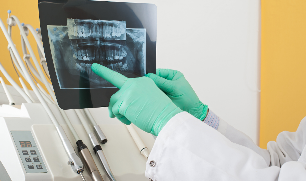
by Ensuring a smooth workflow throughout a dental group can be challenging. Digital radiography offers one solution.
The larger the group practice, the more challenging it can be to get every office on the same page. “Dentists put together protocols to ensure a smooth workflow among their various offices,” says Frank Seipp, product manager, Air Techniques. Many of these protocols are centered on X-ray and digital radiography, he adds. The ability for multiple dental offices to share images and files is an advantage, he says, and companies such as Air Techniques now offer scanners and PSP plates that help make this possible. Dentists working in large group practices today can share one image among their archives, bringing other dentists in the group up to speed on a particular patient case, or they can place the image on a screen to assist with a patient consultation, among other things.
“The advantage of digital radiography is that it doesn’t conform to a single format,” says Seipp. “Digital IPEG images can be passed from one software system to the next and different offices can speak to one another.” Some of Air Techniques’ scanners are designed for chairside use, while other, larger systems are designed for large-volume workflow, he notes. But, all scanners are designed to communicate with the group’s practice management software. Even when offices use different software packages from one another, as long as the images are in standard format, all software systems will accommodate them, he adds.
A sound investment
Converting a group dental practice is an investment – but one worth making, note experts. “Scanners and plates cost tens of thousands of dollars,” says Seipp. “However, when one reviews the time involved using traditional x-ray systems –including labor and maintenance – it’s a worthwhile investment. There are dollars associated with maintaining traditional x-ray equipment, which requires special chemicals and supplies,” he says.
There are several benefits associated with digital radiology. “Digital systems are faster and provide immediate feedback,” Seipp says. Traditional X-rays can slow down the insurance billing process, and can end up delaying the patient’s follow-up with the right specialist and treatment. Also, digital radiography takes up a fraction of the space that traditional X-ray systems do, he says. “There is no need to store the chemicals and supplies associated with traditional X-ray.”
When it comes to digital radiography, there is no one-size-fits-all solution. “Some dental offices prefer using only digital radiography, while others rely on it for specific cases,” says Seipp. “And, some practices still use traditional X-ray.” Dentists and practice managers must consider the goals of the practice, he explains. “What goals is the practice looking to achieve in the near future? There is a wide variety of digital products available and one solution may suit one office’s budget and workflow needs, while another system suits the needs of another office within the practice.” But all digital solutions can communicate with one another, he says, no matter which system each office says.
“The initial setup takes time,” he continues. But, the learning curve is small. And, given the end-of-year Section 179 tax incentives available to dental practices, for some, this might be a good time to move forward.
Choosing the right plate
There is no right or wrong when it comes to selecting a plate to accommodate digital radiographs. Some dentists prefer to work with phosphorous plates, which work much like analog film, while others prefer using hard plate sensors.
Phosphorous plates:
- Digital twain image can be sent to the dentist’s practice management software, archive or laptop, as well as to an insurance agency.
- Available in the same size as traditional X-ray film and, because they are similar to analog film, they are relatively easy for dentists and staff to learn to use.
Hard plate sensors:
- Must be fed into the X-ray machine differently and therefore require about 15 minutes of instruction prior to first-time use.
- The sensors are wired into a PC or laptop.
Shaded Box: How it works
It can be helpful for dentists and their staff to have a basic understanding of how digital radiography works. A chairside digital radiography system produces immediate radiographs in nine seconds or less. The user must simply remove the flexible phosphor sensor from the patient’s mouth, remove the sensor from the infection control barrier envelope and insert it into the slot at the top of the unit. The scanner develops the image, and the sensor is available for reuse.
There is no annual maintenance costs associated with Air Techniques’ ScanX Swift. The system includes sensor cleaning wipes for cleaning the reusable phosphor sensors; a barrier film roll for adhering to the top of the scanner before inserting the PSP and removing between patients; and scanner cleaning sheets for keeping the system’s rollers clean.